Antibody data
- Antibody Data
- Antigen structure
- References [4]
- Comments [0]
- Validations
- Immunohistochemistry [2]
- Flow cytometry [1]
- Blocking/Neutralizing [1]
Submit
Validation data
Reference
Comment
Report error
- Product number
- AF126 - Provider product page

- Provider
- R&D Systems
- Product name
- Human Fas Ligand/TNFSF6 Antibody
- Antibody type
- Polyclonal
- Description
- Antigen Affinity-purified. Detects human Fas Ligand in direct ELISAs and Western blots. In direct ELISAs, approximately 20% cross-reactivity with recombinant mouse Fas Ligand is observed, approximately 7% cross-reactivity with recombinant rat Fas Ligand and recombinant human (rh) BAFF, and less than 1% cross-reactivity with rhTRAIL, rhTNF-alpha , rhGITR Ligand, and rhAPRIL is observed.
- Reactivity
- Human
- Host
- Goat
- Conjugate
- Unconjugated
- Antigen sequence
Q53ZZ1- Isotype
- IgG
- Vial size
- 100 ug
- Concentration
- LYOPH
- Storage
- Use a manual defrost freezer and avoid repeated freeze-thaw cycles. 12 months from date of receipt, -20 to -70 °C as supplied. 1 month, 2 to 8 °C under sterile conditions after reconstitution. 6 months, -20 to -70 °C under sterile conditions after reconstitution.
Submitted references Tissue Inhibitor of Metalloproteinase-3 (TIMP-3) induces FAS dependent apoptosis in human vascular smooth muscle cells.
Activated human CD4+CD45RO+ memory T-cells indirectly inhibit NLRP3 inflammasome activation through downregulation of P2X7R signalling.
Thymosin β10 expression driven by the human TERT promoter induces ovarian cancer-specific apoptosis through ROS production.
Rapid resolution of toxic epidermal necrolysis with anti-TNF-alpha treatment.
English WR, Ireland-Zecchini H, Baker AH, Littlewood TD, Bennett MR, Murphy G
PloS one 2018;13(4):e0195116
PloS one 2018;13(4):e0195116
Activated human CD4+CD45RO+ memory T-cells indirectly inhibit NLRP3 inflammasome activation through downregulation of P2X7R signalling.
Beynon V, Quintana FJ, Weiner HL
PloS one 2012;7(6):e39576
PloS one 2012;7(6):e39576
Thymosin β10 expression driven by the human TERT promoter induces ovarian cancer-specific apoptosis through ROS production.
Kim YC, Kim BG, Lee JH
PloS one 2012;7(5):e35399
PloS one 2012;7(5):e35399
Rapid resolution of toxic epidermal necrolysis with anti-TNF-alpha treatment.
Hunger RE, Hunziker T, Buettiker U, Braathen LR, Yawalkar N
The Journal of allergy and clinical immunology 2005 Oct;116(4):923-4
The Journal of allergy and clinical immunology 2005 Oct;116(4):923-4
No comments: Submit comment
Supportive validation
- Submitted by
- R&D Systems (provider)
- Main image

- Experimental details
- Fas Ligand/TNFSF6 in Human Melanoma. Fas Ligand/TNFSF6 was detected in immersion fixed paraffin-embedded sections of human melanoma tissue using Goat Anti-Human Fas Ligand/TNFSF6 Antigen Affinity-purified Polyclonal Antibody (Catalog # AF126) at 15 µg/mL overnight at 4 °C. Tissue was stained using the Anti-Goat HRP-DAB Cell & Tissue Staining Kit (brown; Catalog # CTS008) and counterstained with hematoxylin (blue). View our protocol for Chromogenic IHC Staining of immersion fixed paraffin-embedded Tissue Sections.
- Submitted by
- R&D Systems (provider)
- Main image

- Experimental details
- Fas Ligand/TNFSF6 in Human Melanoma. Fas Ligand/TNFSF6 was detected in immersion fixed paraffin-embedded sections of human melanoma using Goat Anti-Human Fas Ligand/TNFSF6 Antigen Affinity-purified Polyclonal Antibody (Catalog # AF126) at 15 µg/mL overnight at 4 °C. Tissue was stained using the Anti-Goat HRP-DAB Cell & Tissue Staining Kit (brown; Catalog # CTS008) and counterstained with hematoxylin (blue). Lower panel shows a lack of labeling if primary antibodies are omitted and tissue is stained only with secondary antibody followed by incubation with detection reagents. View our protocol for Chromogenic IHC Staining of Paraffin-embedded Tissue Sections.
Supportive validation
- Submitted by
- R&D Systems (provider)
- Main image

- Experimental details
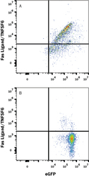
- Detection of Fas Ligand/TNFSF6 in HEK293 Human Cell Line Transfected with Human Fas Ligand/TNFSF6 and eGFP by Flow Cytometry. HEK293 human embryonic kidney cell line transfected with (A) Fas Ligand/TNFSF6 or (B) irrelevant protein, and eGFP were stained with Goat Anti-Human Fas Ligand/TNFSF6 Affinity Purified Polyclonal Antibody (Catalog # AF126) followed by Allophycocyanin-conjugated Anti-Goat IgG Secondary Antibody (Catalog # F0108). Quadrant markers were set based Goat IgG Control Antibody staining (Catalog # AB-108-C, data not shown). View our protocol for Staining Membrane-associated Proteins.
Supportive validation
- Submitted by
- R&D Systems (provider)
- Main image

- Experimental details
- Apoptosis Induced by Fas Ligand/TNFSF6 and Neutralization by Human Fas Ligand/TNFSF6 Antibody. In the presence of a cross-linking antibody, Mouse polyHistidine Monoclonal Antibody (10 µg/mL, Catalog # MAB050), Recombinant Human Fas Ligand/TNFSF6 (Catalog # 126-FL) induces apoptosis in the Jurkat human acute T cell leukemia cell line in a dose-dependent manner (orange line). Apoptosis elicited by Recombinant Human Fas Ligand/TNFSF6 (2 ng/mL) is neutralized (green line) by increasing concentrations of Human Fas Ligand/TNFSF6 Antigen Affinity-purified Polyclonal Antibody (Catalog # AF126). The ND50 is typically 0.12-0.072 μg/mL.
 Explore
 Validate
 Learn
Learn Western blot
 Immunohistochemistry