 Explore
 Validate
 Learn
Learn Western blot
 Immunocytochemistry
Antibody data
- Antibody Data
- Antigen structure
- References [0]
- Comments [0]
- Validations
- Western blot [7]
- Immunocytochemistry [1]
- Immunoprecipitation [1]
- Immunohistochemistry [1]
- Chromatin Immunoprecipitation [1]
Submit
Validation data
Reference
Comment
Report error
- Product number
- GTX110558 - Provider product page

- Provider
- GeneTex
- Proper citation
- GeneTex Cat#GTX110558, RRID:AB_1949671
- Product name
- APE1 antibody
- Antibody type
- Polyclonal
- Reactivity
- Human, Mouse, Rat
- Host
- Rabbit
No comments: Submit comment
Enhanced validation
Supportive validation
- Submitted by
- GeneTex (provider)
- Enhanced method
- Genetic validation
- Main image

- Experimental details
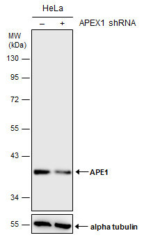
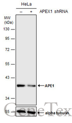
- Non-transfected (¡V) and transfected (+) HeLa whole cell extracts (30 ?g) were separated by 10% SDS-PAGE, and the membrane was blotted with APE1 antibody (GTX110558) diluted at 1:1000. The HRP-conjugated anti-rabbit IgG antibody (GTX213110-01) was used to detect the primary antibody.
Supportive validation
- Submitted by
- GeneTex (provider)
- Main image
- Experimental details
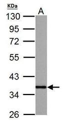
- APE1 antibody detects APEX1 protein by western blot analysis.A. 30 ?g PC-12 whole cell lysate/extract10% SDS-PAGEAPE1 antibody (GTX110558) dilution: 1:10000 The HRP-conjugated anti-rabbit IgG antibody (GTX213110-01) was used to detect the primary antibody.
- Submitted by
- GeneTex (provider)
- Main image

- Experimental details
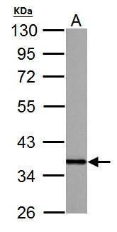
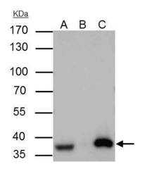
- Sample (30 ug of whole cell lysate) A: A431 (GTX27909) B: Hela C: Molt-4 (GTX27912) 10% SDS PAGE GTX110558 diluted at 1:1000
- Submitted by
- GeneTex (provider)
- Main image
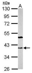
- Experimental details
- Sample (50 ug of whole cell lysate) A: mouse brain 10% SDS PAGE GTX110558 diluted at 1:2000

- Submitted by
- GeneTex (provider)
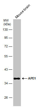
- Main image
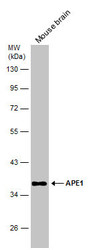
- Experimental details
- Mouse tissue extract (50 ?g) was separated by 10% SDS-PAGE, and the membrane was blotted with APE1 antibody (GTX110558) diluted at 1:1000. The HRP-conjugated anti-rabbit IgG antibody (GTX213110-01) was used to detect the primary antibody.
- Submitted by
- GeneTex (provider)
- Main image
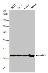
- Experimental details
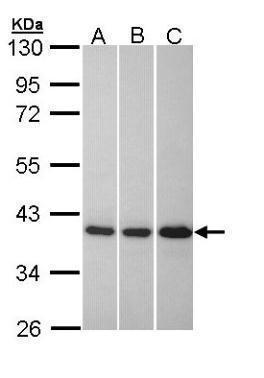
- Various whole cell extracts (30 ?g) were separated by 10% SDS-PAGE, and the membrane was blotted with APE1 antibody (GTX110558) diluted at 1:5000. The HRP-conjugated anti-rabbit IgG antibody (GTX213110-01) was used to detect the primary antibody.

- Submitted by
- GeneTex (provider)
- Main image
- Experimental details
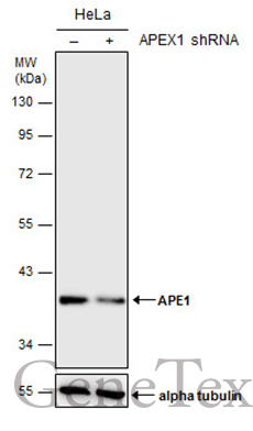
- Non-transfected (¡V) and transfected (+) HeLa whole cell extracts (30 ?g) were separated by 10% SDS-PAGE, and the membrane was blotted with APE1 antibody (GTX110558) diluted at 1:1000. The HRP-conjugated anti-rabbit IgG antibody (GTX213110-01) was used to detect the primary antibody.
Supportive validation
- Submitted by
- GeneTex (provider)
- Main image
- Experimental details
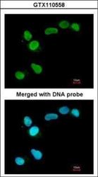
- Immunofluorescence analysis of paraformaldehyde-fixed A549, using APE1(GTX110558) antibody at 1:200 dilution.

Supportive validation
- Submitted by
- GeneTex (provider)
- Main image
- Experimental details
- APE1 antibody immunoprecipitates APE1 protein in IP experiments. IP Sample: A431 whole cell lysate/extract A : 30 £gg whole cell lysate/extract of APE1 protein expressing A431 cells B : Control with 3 £gg of pre-immune rabbit IgG C : Immunoprecipitation of APE1 by 3 £gg of APE1 antibody (GTX110558) 10% SDS-PAGE The immunoprecipitated APE1 protein was detected by APE1 antibody (GTX110558) diluted at 1 : 1000. EasyBlot anti-rabbit IgG (HRP) (GTX221666-01) was used as a secondary reagent.

Supportive validation
- Submitted by
- GeneTex (provider)
- Main image

- Experimental details
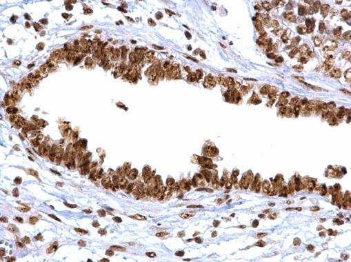
- APE1 antibody detects APE1 protein at cytoplasm, nucleus and nucleolus on human ovarian carcinoma by immunohistochemical analysis. Sample: Paraffin-embedded human ovarian carcinoma. APE1 antibody (GTX110558) diluted at 1:500.
Supportive validation
- Submitted by
- GeneTex (provider)
- Main image
- Experimental details
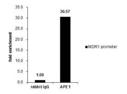
- Cross-linked ChIP was performed with 293T chromatin extract treated with Trichostatin A (0.4 £gM for 18 h) and 5 £gg of either control rabbit IgG or anti-APE1 antibody. The precipitated DNA was detected by PCR with primer set targeting to MDR1 promoter.
