 Explore
 Validate
 Learn
LearnPA1-16500
antibody from Invitrogen Antibodies
Targeting: APEX1
APE, APE-1, APEN, APEX, APX, HAP1, REF-1, REF1
 Western blot Immunocytochemistry
 Immunoprecipitation Immunohistochemistry Flow cytometry Blocking/Neutralizing Chromatin Immunoprecipitation
Antibody data
- Antibody Data
- Antigen structure
- References [0]
- Comments [0]
- Validations
- Western blot [5]
- Immunocytochemistry [2]
- Immunohistochemistry [1]
Submit
Validation data
Reference
Comment
Report error
- Product number
- PA1-16500 - Provider product page

- Provider
- Invitrogen Antibodies
- Product name
- APE1 Polyclonal Antibody
- Antibody type
- Polyclonal
- Antigen
- Purifed from natural sources
- Description
- In IHC applications, this antibody can be competitively inhibited from recognizing the APE1 antigen in tissues using APE1 protein. This antibody can be used on frozen sections, fixed-paraffin sections and cytospin preps. Suggested positive control: Hela whole cell extract, antigen standard for APEX1 (transient overexpression lysate).
- Reactivity
- Human, Mouse, Rat, Rabbit
- Host
- Rabbit
- Isotype
- IgG
- Vial size
- 100 µL
- Concentration
- 1 mg/mL
- Storage
- -20° C, Avoid Freeze/Thaw Cycles
No comments: Submit comment
Supportive validation
- Submitted by
- Invitrogen Antibodies (provider)
- Main image

- Experimental details
- Western blot in HeLa whole cell extract using Product # PA1-16500.

- Submitted by
- Invitrogen Antibodies (provider)
- Main image

- Experimental details
- Western blot analysis of APE1 in cell lysates of 1. HeLa, 2. Ntera2, 3. A431, 4. HepG2, 5. MCF7, 6. NIH 3T3, 7. PC12, and 8. Cos 7. Samples were incubated in APE1 polyclonal antibody (Product # PA1-16500).

- Submitted by
- Invitrogen Antibodies (provider)
- Main image

- Experimental details
- Western blot analysis of APE1 in 0.1 mg/mL HeLa lysate. Samples were incubated in APE1 polyclonal (Product # PA1-16500). This experiment was performed under reducing conditions using the 12-230 kDa separation system. * Non-specific interaction with the 230 kDa Simple Western standard may be seen with this antibody.

- Submitted by
- Invitrogen Antibodies (provider)
- Main image

- Experimental details
- Knockdown of APE1 was achieved by transfecting HeLa with APE1 specific siRNAs (Silencer® select Product # s1446, s1445). Western blot analysis (Fig. a) was performed using Nuclear enriched extracts from the APE1 knockdown cells (lane 3), non-targeting scrambled siRNA transfected cells (lane 2) and untransfected cells (lane 1). The blot was probed with APE1 Polyclonal Antibody (Product # PA1-16500, 1:1000 dilution ) and Goat anti-Rabbit IgG (H+L) Superclonal™ Recombinant Secondary Antibody, HRP (Product # A27036, 1:4000). Densitometric analysis of this western blot is shown in histogram (Fig. b). Decrease in signal upon siRNA mediated knock down confirms that antibody is specific to APE1.

- Submitted by
- Invitrogen Antibodies (provider)
- Main image

- Experimental details
- Western blot was performed using Anti-APE1 Polyclonal Antibody(Product # PA1-16500) and a 30 kDa band corresponding to APE1 was observed across cell lines and tissues tested. Nuclear enriched extracts (30µg lysate) of HeLa (Lane 1), PC-3 (Lane 2), A549 (Lane 3), A-431(Lane 4), HT-29 (Lane 5), Mouse Liver (Lane 6) and Rat Liver (Lane 7) were electrophoresed using NuPAGE™ 4-12% Bis-Tris Protein Gel (Product # NP0322BOX). Resolved proteins were then transferred onto a Nitrocellulose membrane (Product # LC2002) by iBlot® 2 Dry Blotting System (Product # IB21001). The blot was probed with the primary antibody (1:1000 Dilution) and detected by chemiluminescence with Goat anti-Rabbit IgG (H+L) Superclonal™ Recombinant Secondary Antibody, HRP (Product # A27036,1:4000 dilution) using the iBright FL 1000 (Product # A32752). Chemiluminescent detection was performed using Novex® ECL Chemiluminescent Substrate Reagent Kit (Product # WP20005).

Supportive validation
- Submitted by
- Invitrogen Antibodies (provider)
- Main image
- Experimental details
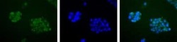
- Immunocytochemistry analysis of APE1 in HepG2 cells. Samples were incubated in APE1 polyclonal antibody (Product # PA1-16500). APE1 (Green). Nuclei (Blue) are counterstained with Hoechst 33258.

- Submitted by
- Invitrogen Antibodies (provider)
- Main image

- Experimental details
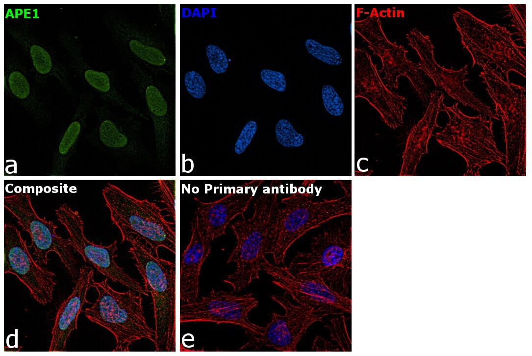
- Immunofluorescence analysis of APE1 was performed using 70% confluent log phase HeLa cells. The cells were fixed with 4% paraformaldehyde for 10 minutes, permeabilized with 0.1% Triton™ X-100 for 15 minutes, and blocked with 2% BSA for 45 minutes at room temperature. The cells were labeled with APE1 Polyclonal Antibody (Product # PA1-16500) at 1:100 Dilution in 0.1% BSA, incubated at 4 degree celsius overnight and then labeled with Goat anti-Rabbit IgG (H+L) Superclonal™ Recombinant Secondary Antibody, Alexa Fluor® 488 conjugate (Product # A27034), (1:2000), for 45 minutes at room temperature (Panel a: Green). Nuclei (Panel b:Blue) were stained with SlowFade® Gold Antifade Mountant with DAPI (Product # S36938). F-actin (Panel c: Red) was stained with Rhodamine Phalloidin (Product # R415, 1:300 dilution). Panel d represents the merged image showing Nucleus localization. Panel e represents control cells with no primary antibody to assess background. The images were captured at 60x magnification.
Supportive validation
- Submitted by
- Invitrogen Antibodies (provider)
- Main image

- Experimental details
- Immunohistochemical analysis of APE1 in prostate cancer. Samples were incubated in APE1 polyclonal antibody (Product # PA1-16500).
