Explore
 Validate
 Learn
Learn Western blot
Antibody data
- Antibody Data
- Antigen structure
- References [1]
- Comments [0]
- Validations
- Western blot [1]
Submit
Validation data
Reference
Comment
Report error
- Product number
- MAB1044 - Provider product page

- Provider
- R&D Systems
- Product name
- Human/Mouse/Rat APE Antibody
- Antibody type
- Monoclonal
- Description
- Protein A or G purified from hybridoma culture supernatant. Detects human, mouse and rat APE in Western blots.
- Reactivity
- Human, Mouse, Rat
- Host
- Mouse
- Conjugate
- Unconjugated
- Antigen sequence
P27695- Isotype
- IgG
- Antibody clone number
- 200913
- Vial size
- 100 ug
- Concentration
- LYOPH
- Storage
- Use a manual defrost freezer and avoid repeated freeze-thaw cycles. 12 months from date of receipt, -20 to -70 °C as supplied. 1 month, 2 to 8 °C under sterile conditions after reconstitution. 6 months, -20 to -70 °C under sterile conditions after reconstitution.
Submitted references Characterization of Gaucher disease bone marrow mesenchymal stromal cells reveals an altered inflammatory secretome.
Campeau PM, Rafei M, Boivin MN, Sun Y, Grabowski GA, Galipeau J
Blood 2009 Oct 8;114(15):3181-90
Blood 2009 Oct 8;114(15):3181-90
No comments: Submit comment
Supportive validation
- Submitted by
- R&D Systems (provider)
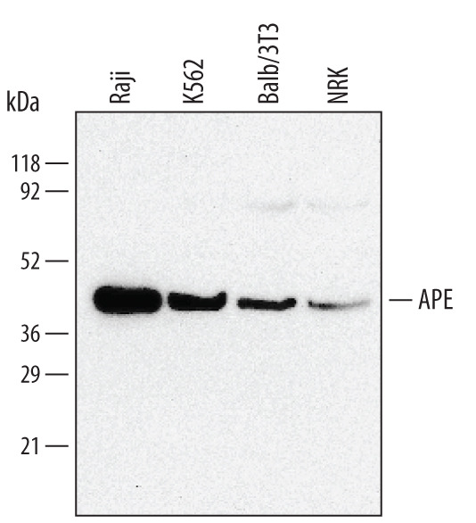
- Main image
- Experimental details
- Detection of Human/Mouse/Rat APE by Western Blot. Western blot shows lysates of NRK rat normal kidney cell line, Raji human Burkitt's lymphoma cell line, K562 human chronic myelogenous leukemia cell line, and Balb/3T3 mouse embryonic fibroblast cell line. PVDF membrane was probed with 0.5 µg/mL of Human/Mouse/Rat APE Monoclonal Antibody (Catalog # MAB1044) followed by HRP-conjugated Anti-Mouse IgG Secondary Antibody (Catalog # HAF007). A specific band was detected for APE at approximately 40 kDa (as indicated). This experiment was conducted under reducing conditions and using Immunoblot Buffer Group 1.