 Explore
 Validate
 Learn
LearnPA5-101023
antibody from Invitrogen Antibodies
Targeting: PMEL
D12S53E, gp100, Pmel17, SI, SIL, SILV
 Western blot
Antibody data
- Antibody Data
- Antigen structure
- References [1]
- Comments [0]
- Validations
- Western blot [2]
- Immunocytochemistry [1]
- Immunohistochemistry [1]
- Other assay [1]
Submit
Validation data
Reference
Comment
Report error
- Product number
- PA5-101023 - Provider product page

- Provider
- Invitrogen Antibodies
- Product name
- PMEL Polyclonal Antibody
- Antibody type
- Polyclonal
- Antigen
- Synthetic peptide
- Reactivity
- Human, Mouse
- Host
- Rabbit
- Isotype
- IgG
- Vial size
- 100 µL
- Concentration
- 1 mg/mL
- Storage
- -20°C
Submitted references Comparison of human amniotic membrane decellularisation approaches for hESC-derived RPE cells culture.
Daniele E, Ferrari B, Rassu N, Ben-Nun J, Bosio L, Barbaro V, Ferrari S, Ponzin D
BMJ open ophthalmology 2022 Sep;7(1)
BMJ open ophthalmology 2022 Sep;7(1)
No comments: Submit comment
Supportive validation
- Submitted by
- Invitrogen Antibodies (provider)
- Main image
- Experimental details
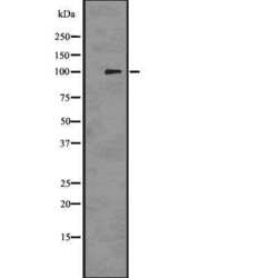
- Western blot analysis of PMEL in 293 cell lysate. Samples were incubated with PMEL polyclonal antibody (Product # PA5-101023).

- Submitted by
- Invitrogen Antibodies (provider)
- Main image
- Experimental details
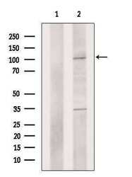
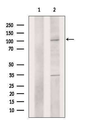
- Western blot analysis of PMEL in 3T3 cell lysate (left lane: treated with blocking peptide). Samples were incubated with PMEL polyclonal antibody (Product # PA5-101023).
Supportive validation
- Submitted by
- Invitrogen Antibodies (provider)
- Main image
- Experimental details
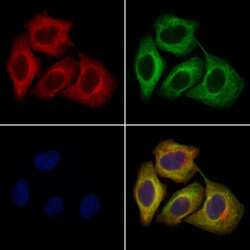
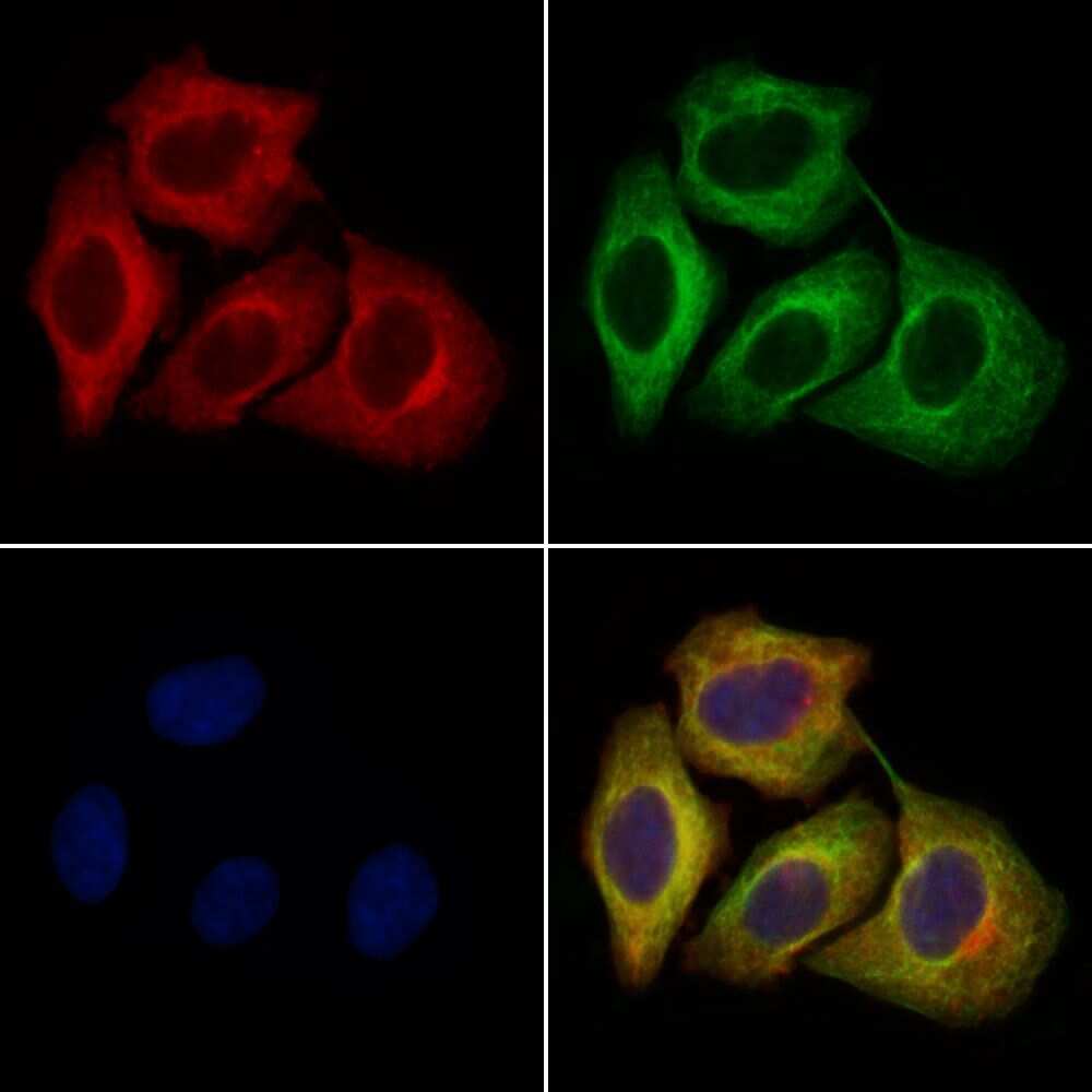
- Immunofluorescent analysis of PMEL in HeLa cells. Samples were fixed with paraformaldehyde, permeabilized with 0.1% Triton X-100, blocked with 10% serum (45 min at 25°C), incubated with mouse anti-beta tubulin and PMEL polyclonal antibody (Product # PA5-101023) using a dilution of 1:200 (1 hr, 37°C), and followed by goat anti-rabbit IgG Alexa Fluor 594 (red) and goat anti-mouse IgG Alexa Fluor 488 (green).
Supportive validation
- Submitted by
- Invitrogen Antibodies (provider)
- Main image
- Experimental details
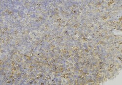
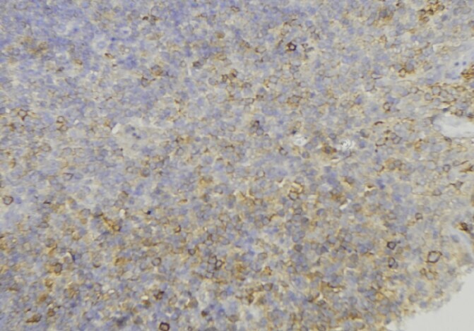
- Immunohistochemistry analysis of paraffin-embedded PMEL in human lymph node tissue. Antigen retrieval was performed using citrate buffer. Samples were blocked with blocking buffer (1.5 hr, 22°C), incubated with PMEL polyclonal antibody (Product # PA5-101023) using a dilution of 1:100 (1.5 hr, 22°C), followed by HRP conjugated goat anti-rabbit.
Supportive validation
- Submitted by
- Invitrogen Antibodies (provider)
- Main image
- Experimental details
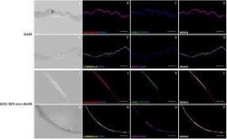
- Immunofluorescent staining of RPE differentiation markers, PMEL-17 (C, K; green) and RPE65 (G, O; magenta) and BM markers, type IV collagen (B, J; red) and laminin alpha5 (F, N; yellow) in cryopreserved ihAM (A-H) and hESC-RPE cells culture on dhAM following thermolysin treatment (I-P). RPE markers identified differentiated hESC-RPE cells over dhAM (K, O), while BM markers demonstrated the preservation of the BM following thermolysin treatment (J, N). Merged images showed areas of membrane where RPE markers were not detected, despite a proper distribution of the BM markers (L, P). ihAM was used as control. hAECs, RPE cells and stromal cells nuclei were stained with DAPI (blue). Scale bars=100 um. BM, basement membrane; dhAM, denuded hAM; hAM, human amniotic membrane; hESC, human embryonic stem cell; ihAM, intact hAM; RPE, retinal pigment epithelium.
