 Explore
 Validate
 Learn
Learn Western blot
 Immunohistochemistry
Antibody data
- Antibody Data
- Antigen structure
- References [1]
- Comments [0]
- Validations
- Western blot [2]
- Immunocytochemistry [2]
- Immunohistochemistry [6]
Submit
Validation data
Reference
Comment
Report error
- Product number
- HPA002087 - Provider product page

- Provider
- Atlas Antibodies
- Proper citation
- Atlas Antibodies Cat#HPA002087, RRID:AB_1079245
- Product name
- Anti-LEF1
- Antibody type
- Polyclonal
- Reactivity
- Human
- Host
- Rabbit
- Conjugate
- Unconjugated
- Antigen sequence
SDVNSKQGMSRHPPAPDIPTFYPLSPGGVGQITPP
LGWQGQPVYPITGGFRQPYPSSLSVDTSMSRFSHH
MIPGPPGPHTTGIPHPAIVTPQVKQEHPHTDSDLM
HVKPQHEQRKEQEPKRPHIKKPLNAFMLY- Isotype
- IgG
- Vial size
- 100 µl
- Storage
- Store at +4°C for short term storage. Long time storage is recommended at -20°C.
Submitted references Expression profiling of microdissected cell populations selected from basal cells in normal epidermis and basal cell carcinoma
Asplund A, Gry Björklund M, Sundquist C, Strömberg S, Edlund K, Östman A, Nilsson P, Pontén F, Lundeberg J
British Journal of Dermatology 2008 March;158(3):527-538
British Journal of Dermatology 2008 March;158(3):527-538
No comments: Submit comment
Supportive validation
Supportive validation
- Submitted by
- Atlas Antibodies (provider)
- Enhanced method
- Orthogonal validation
- Main image
- Experimental details
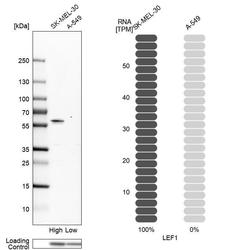
- Western blot analysis in human cell lines SK-MEL-30 and A-549 using Anti-LEF1 antibody. Corresponding LEF1 RNA-seq data are presented for the same cell lines. Loading control: Anti-PPIB.

Supportive validation
- Submitted by
- Atlas Antibodies (provider)
- Enhanced method
- Recombinant expression validation
- Main image
- Experimental details
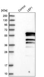
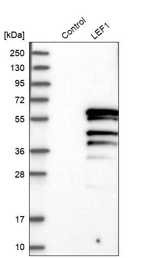
- Western blot analysis in control (vector only transfected HEK293T lysate) and LEF1 over-expression lysate (Co-expressed with a C-terminal myc-DDK tag (~3.1 kDa) in mammalian HEK293T cells, LY402529).
Supportive validation
- Submitted by
- Atlas Antibodies (provider)
- Main image

- Experimental details
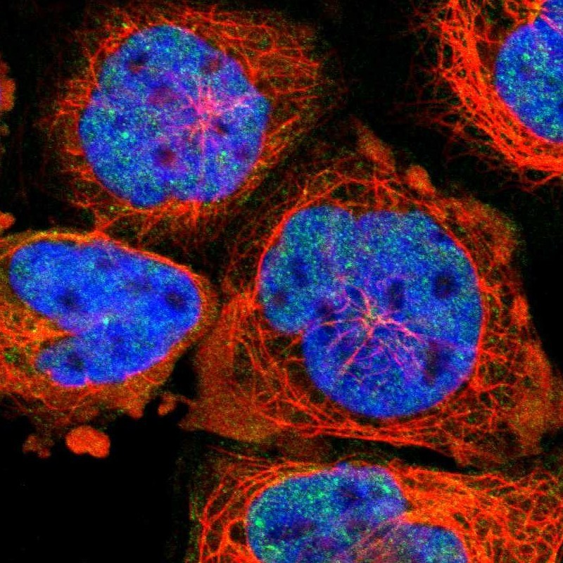
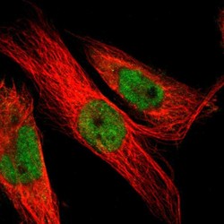
- Immunofluorescent staining of human cell line A-431 shows localization to nucleoplasm, nuclear bodies & cytosol.
- Submitted by
- Atlas Antibodies (provider)
- Main image
- Experimental details
- Immunofluorescent staining of human cell line U-251 MG shows localization to nucleoplasm & cytosol.
- Sample type
- HUMAN

Enhanced validation
Supportive validation
- Submitted by
- Atlas Antibodies (provider)
- Enhanced method
- Orthogonal validation
- Main image
- Experimental details
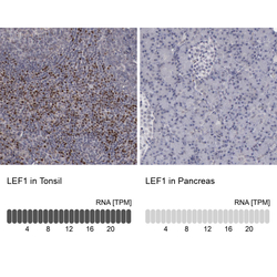
- Immunohistochemistry analysis in human tonsil and pancreas tissues using HPA002087 antibody. Corresponding LEF1 RNA-seq data are presented for the same tissues.
- Sample type
- HUMAN

Supportive validation
- Submitted by
- Atlas Antibodies (provider)
- Main image

- Experimental details
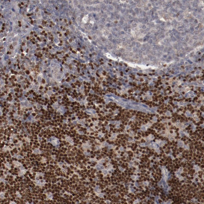
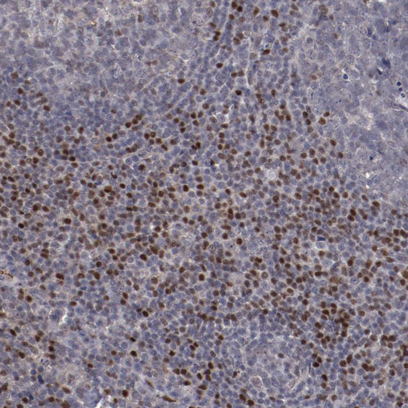
- Immunohistochemical staining of human lymph node shows strong nuclear positivity in non-germinal center cells.
- Submitted by
- Atlas Antibodies (provider)
- Main image
- Experimental details
- Immunohistochemical staining of human lymph node shows moderate to strong nuclear positivity in non - germinal center cells.
- Sample type
- HUMAN

- Submitted by
- Atlas Antibodies (provider)
- Main image

- Experimental details
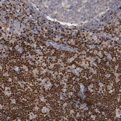
- Immunohistochemical staining of human tonsil shows moderate nuclear positivity in non - germinal center cells.
- Sample type
- HUMAN
- Submitted by
- Atlas Antibodies (provider)
- Main image
- Experimental details
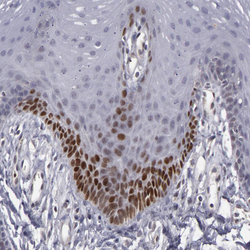
- Immunohistochemical staining of human oral mucosa shows moderate nuclear positivity in subset of squamous epithelial cells.
- Sample type
- HUMAN

- Submitted by
- Atlas Antibodies (provider)
- Main image
- Experimental details
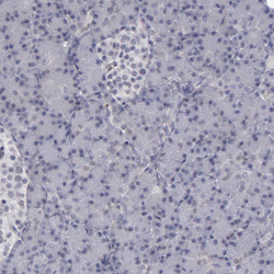
- Immunohistochemical staining of human pancreas shows no nuclear positivity as expected.
- Sample type
- HUMAN
