Explore
 Validate
 Learn
Learn Western blot
Antibody data
- Antibody Data
- Antigen structure
- References [0]
- Comments [0]
- Validations
- Western blot [2]
- Immunocytochemistry [2]
- Immunohistochemistry [4]
- Flow cytometry [2]
Submit
Validation data
Reference
Comment
Report error
- Product number
- MA5-36233 - Provider product page

- Provider
- Invitrogen Antibodies
- Product name
- CFL2 Monoclonal Antibody (8C13)
- Antibody type
- Monoclonal
- Antigen
- Synthetic peptide
- Description
- Reconstitute with 0.2 mL of distilled water to yield a concentration of 500 µg/mL.
- Reactivity
- Human, Mouse, Rat
- Host
- Mouse
- Isotype
- IgG
- Antibody clone number
- 8C13
- Vial size
- 100 µg
- Concentration
- 500 µg/mL
- Storage
- Store at 4°C short term. For long term storage, store at -20°C, avoiding freeze/thaw cycles.
No comments: Submit comment
Supportive validation
- Submitted by
- Invitrogen Antibodies (provider)
- Main image

- Experimental details
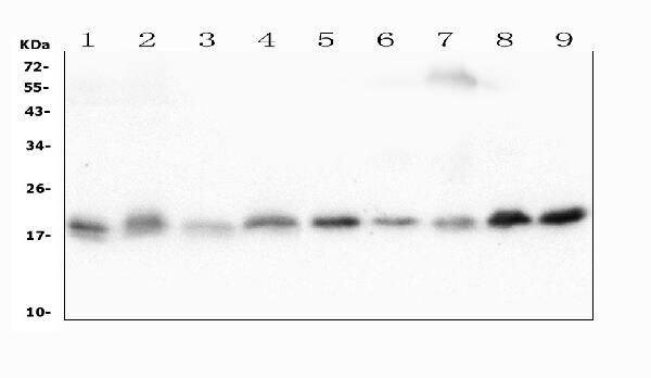
- Western blot analysis of CFL2 in the following samples: Lane 1: rat heart tissue lysates, Lane 2: rat liver tissue lysates, Lane 3: rat kidney tissue lysates, Lane 4: rat brain tissue lysates, Lane 5: mouse heart tissue lysates, Lane 6: mouse liver tissue lysates, Lane 7: mouse kidney tissue lysates, Lane 8: mouse brain tissue lysates, Lane 9: mouse NIH/3T3 whole cell lysates. Samples consisting of 50 µg (reducing conditions) of protein was separated with 5-20% SDS-PAGE gel (70V, Stacking gel; 90V, Resolving gel; 2-3 hrs.), transferred to a Nitrocellulose membrane (150mA, 50-90 min) and washed with TBS-0.1% Tween (3 times, 5 minutes/wash) and blocked with 5% Non-fat Milk/TBS (1.5 hrs., room temperature). The membrane was incubated in CFL2 monoclonal antibody (Product # MA5-36233) at a dilution of 0.5 µg/mL (overnight, 4°C), followed by goat anti-mouse IgG-HRP and chemiluminescence (ECL) with a dilution of 1:10,000 (1.5 hours, room temperature).
- Submitted by
- Invitrogen Antibodies (provider)
- Main image

- Experimental details
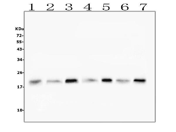
- Western blot analysis of CFL2 in the following samples: Lane 1: human HeLA whole cell lysates, Lane 2: human U2OS whole cell lysates, Lane 3: human HepG2 whole cell lysates, Lane 4: human T-47D whole cell lysates, Lane 5: human Raji whole cell lysates, Lane 6: human placenta tissue lysates, Lane 7: human A549 whole cell lysates. Samples consisting of 50 µg (reducing conditions) of protein was separated with 5-20% SDS-PAGE gel (70V, Stacking gel; 90V, Resolving gel; 2-3 hrs.), transferred to a Nitrocellulose membrane (150mA, 50-90 min) and washed with TBS-0.1% Tween (3 times, 5 minutes/wash) and blocked with 5% Non-fat Milk/TBS (1.5 hrs., room temperature). The membrane was incubated in CFL2 monoclonal antibody (Product # MA5-36233) at a dilution of 0.5 µg/mL (overnight, 4°C), followed by goat anti-mouse IgG-HRP and chemiluminescence (ECL) with a dilution of 1:10,000 (1.5 hours, room temperature).
Supportive validation
- Submitted by
- Invitrogen Antibodies (provider)
- Main image

- Experimental details
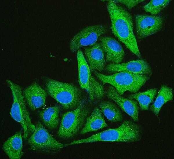
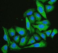
- Immunocytochemistry analysis of CFL2 in U20S cell. Antigen retrieval was performed with enzyme antigen retrieval (15 min). Samples were blocked with 10% goat serum and incubated in CFL2 monoclonal antibody (Product # MA5-36233) at a dilution of 2 µg/mL (overnight, 4°C), followed by DyLight 488 conjugated goat anti-mouse IgG (30 min, 37°C) and DAPI at a dilution of 1:100.
- Submitted by
- Invitrogen Antibodies (provider)
- Main image
- Experimental details
- Immunocytochemistry analysis of CFL2 in U20S cell. Antigen retrieval was performed with enzyme antigen retrieval (15 min). Samples were blocked with 10% goat serum and incubated in CFL2 monoclonal antibody (Product # MA5-36233) at a dilution of 2 µg/mL (overnight, 4°C), followed by DyLight 488 conjugated goat anti-mouse IgG (30 min, 37°C) and DAPI at a dilution of 1:100.

Supportive validation
- Submitted by
- Invitrogen Antibodies (provider)
- Main image

- Experimental details
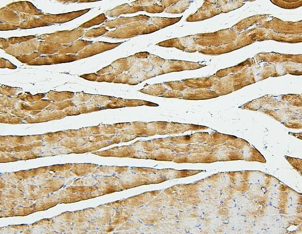
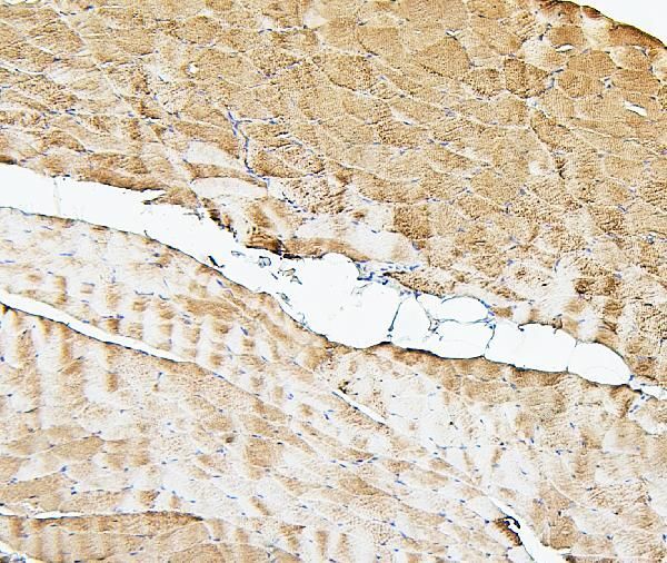
- Immunohistochemistry analysis of CFL2 in paraffin-embedded rat skeletal muscle tissues. Antigen retrieval was performed with citrate buffer (pH6, 20 min). Samples were blocked with 10% goat serum and incubated in CFL2 monoclonal antibody (Product # MA5-36233) at a dilution of 1 µg/mL (overnight, 4°C), followed by biotinylated goat anti-mouse IgG (30 min, 37°C) and Strepavidin-Biotin-Complex (SABC) with DAB.
- Submitted by
- Invitrogen Antibodies (provider)
- Main image
- Experimental details
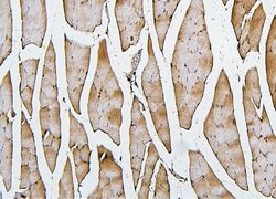
- Immunohistochemistry analysis of CFL2 in paraffin-embedded mouse skeletal muscle tissues. Antigen retrieval was performed with citrate buffer (pH6, 20 min). Samples were blocked with 10% goat serum and incubated in CFL2 monoclonal antibody (Product # MA5-36233) at a dilution of 1 µg/mL (overnight, 4°C), followed by biotinylated goat anti-mouse IgG (30 min, 37°C) and Strepavidin-Biotin-Complex (SABC) with DAB.

- Submitted by
- Invitrogen Antibodies (provider)
- Main image
- Experimental details
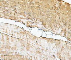
- Immunohistochemistry analysis of CFL2 in paraffin-embedded human skeletal muscle tissues. Antigen retrieval was performed with citrate buffer (pH6, 20 min). Samples were blocked with 10% goat serum and incubated in CFL2 monoclonal antibody (Product # MA5-36233) at a dilution of 1 µg/mL (overnight, 4°C), followed by biotinylated goat anti-mouse IgG (30 min, 37°C) and Strepavidin-Biotin-Complex (SABC) with DAB.
- Submitted by
- Invitrogen Antibodies (provider)
- Main image
- Experimental details
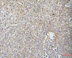
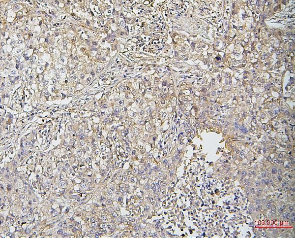
- Immunohistochemistry analysis of CFL2 in paraffin-embedded human lung cancer tissues. Antigen retrieval was performed with citrate buffer (pH6, 20 min). Samples were blocked with 10% goat serum and incubated in CFL2 monoclonal antibody (Product # MA5-36233) at a dilution of 1 µg/mL (overnight, 4°C), followed by biotinylated goat anti-mouse IgG (30 min, 37°C) and Strepavidin-Biotin-Complex (SABC) with DAB.
Supportive validation
- Submitted by
- Invitrogen Antibodies (provider)
- Main image
- Experimental details
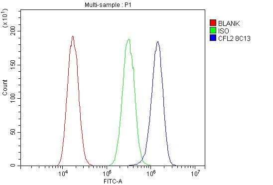
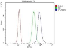
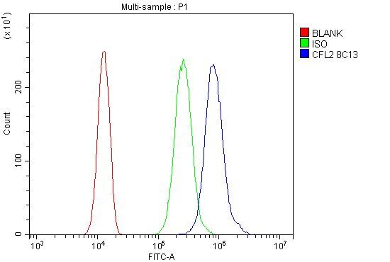
- Flow cytometry analysis of CFL2 in SiHa cells. Samples were blocked with 10% normal goat serum and incubated in CFL2 monoclonal antibody (Product # MA5-36233) at a dilution of 1 µg/1x10^6 cells (30 min, 20°C), followed by Dylight 488 conjugated goat anti-mouse IgG (30 min, 37°C) with a dilution of 5-10 µg/1x10^6 cells (30 min, 20°C). Isotype control antibody (Green line) was mouse IgG (1 µg/1x10^6) and unlabeled sample (Red line).
- Submitted by
- Invitrogen Antibodies (provider)
- Main image
- Experimental details
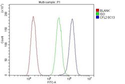
- Flow cytometry analysis of CFL2 in A549 cells. Samples were blocked with 10% normal goat serum and incubated in CFL2 monoclonal antibody (Product # MA5-36233) at a dilution of 1 µg/1x10^6 cells (30 min, 20°C), followed by Dylight 488 conjugated goat anti-mouse IgG (30 min, 37°C) with a dilution of 5-10 µg/1x10^6 cells (30 min, 20°C). Isotype control antibody (Green line) was mouse IgG (1 µg/1x10^6) and unlabeled sample (Red line).